
Radiology - Imaging Services 
Imaging Services at Glendive Medical Center include:
General Radiography - the use of radiation (x-rays) to produce black and white images of anatomy. X-rays may be used to detect bone fractures, find foreign objects in the body, and demonstrate the relationship between bone and soft tissue. The most common type of x-ray exam is chest radiography.
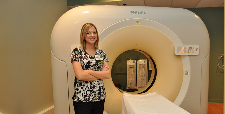
Computed Tomography (CT) - the use of a rotating x-ray unit to obtain "slices" of anatomy at different levels within the body. A computer then stacks and assembles the individual slices, creating a diagnostic image. With CT technology, physicians can view the inside of organs, a feat not possible with general radiography.
Ultrasound - the use of sound waves to obtain images of organs and tissues in the body. During an ultrasound examination, the sonographer (ultrasound technologist) places a transducer in contact wit the patient's body. It emits high-frequency sound waves that pass through the body, sending back "echoes" as they bounce off organs and tissues. Special computer equipment converts those echoes into visual data.

Digital Mammography - also called full-field digital mammography (FFDM), is a mammography system in which the x-ray film is replaced by detectors, similar to those found in digital cameras, that convert x-rays into electrical signals. These electrical signals are then used to produce images of the breast that can be seen on a computer screen or printed on special film.
Bone Densitometry - the use of special type of x-ray equipment to measure bone mineral density at specific anatomical sites. These results can be used by physicians to estimate the amount of bone loss due to osteoporosis, to track the rate of bone loss over a specific period of time, and to estimate the risk of fracture.
Magnetic Resonance Imaging (MRI) - During a MRI exam, the atoms in the patient's body are exposed to a strong magnetic field. The technologist applies a radiofrequency pulse to the field, which knocks the atoms out of alignment. When the technologist turns the pulse off, the atoms return to their original position. In the process, they give off signals that are measured by a computer and processed to create detailed images of the patient's anatomy.
Radiologic technologists (radiographers) are the medical personnel who perform diagnostic imaging examinations and are responsible for accurately positioning patients and ensuring that a quality diagnostic image is produced. They are educated in anatomy, patient position, examination techniques, equipment protocols, radiation safety, radiation protection and basic patient care. All radiologic technologists employed at GMC are licensed by the State of Montana Board of Radiologic Technologists.
Radiologists are physicians who specialize in the interpretation of medical images. They are specially trained to identify injury and disease in each of the body's systems. Radiologists earn a four-year doctoral degree to become an M.D. (medical doctor) or D.O. (doctor of osteopathy). They then complete a four-year residency in diagnostic radiology. All radiologists employed at GMC are licensed by the State of Montana Board of Medical Examiners.